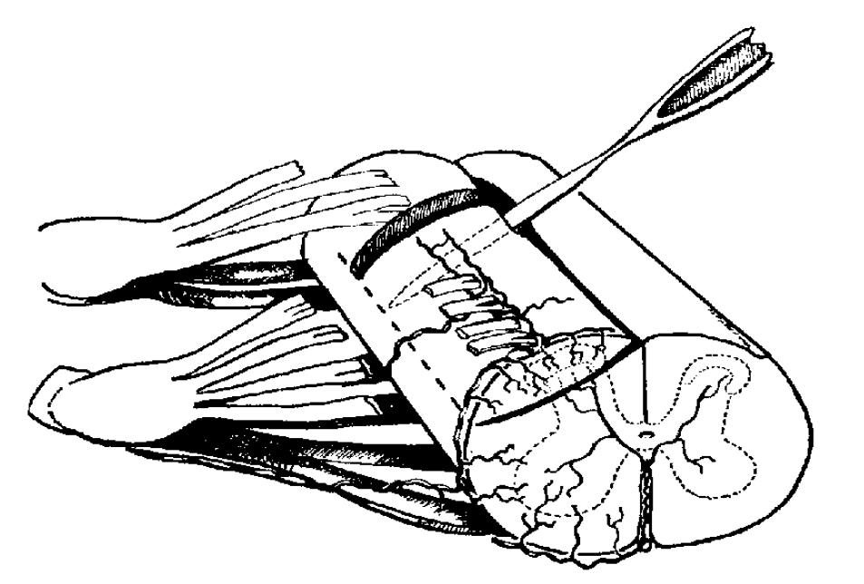
Diagram illustrating the operative procedure used for preparing an „isolated dorsal column”. By not cutting through the pial surface at the lateral side of the cord (along the dashed line) the arcuate vessels are spared for blood supply of the substantia gelatinosa.
Having experienced the power of the silver technique, Szentágothai decided to break up the homogenously looking gelatinous substance in the dorsal tip of the spinal gray matter, laminae I,II and III. He combined the secondary fibre and synapse degeneration technique with the Golgi method and extended to explore the form of neurons in this terra incognita. He had become aware of the difficulty caused by the haze of innumerable neural elements heaped up in this small circumscribed space, and introduced the isolated dorsal horn stratagem.
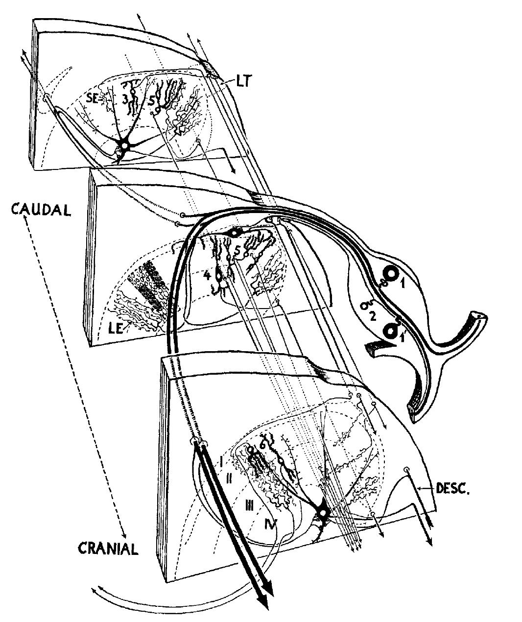
Semi-schematic drawing of the left dorsal sector of the spinal cord with three successice levels, less than one segment apart, to illustrate neuronal arrangement in the substantia gelatinosa. Cytoarchitectonical layers according to Rexed (’54) are indicated by roman numerals. (1) and (1’) large calibered cutaneous afferents, (1) projecting from the dorsal part of the dermatome and (1’) the ventral ramus with their central connections of large endings (LE) in the substantia gelatinosa (SG) and boutons-terminaux on large neurons of lamina IV. (2) Small calibered cutaneous afferent with axon turning into the medial part of Lissauuer’s tract (LT) and giving small endings (SE) to the SG as well as axosomatic knobs to large marginal neurons of lamina I (middle section). (3) SG neurons proper that send their axon into the lateral division of LT, (4) larger SG neurons sending their axons into the lateral fasciculus proprius. Axons of both types terminate again in the SG by small terminals. (5) SG neurons giving their axon to its longitudinal axonal system and establishing synapses with the dendrites of the large lamina IV neurons (cranial section at bottom figure.) (6) SG neurons with axons crossing in the posterior commissure to contralateral SG. DESC = descending mainly pyramidal fibers that terminate with boutons-terminaux on large lamina neurons. Larger rectangle in middle section indicates the relation of the distribution field of large primary sensory terminals and size and density of cell bodies in the SG, the smaller hatched rectangles indicate average size of distribution field of small primary sensory terminals.
With a dorso-ventral oriented oblique cut and two vertical cuts (one rostral and another caudal) inserted into the spinal cord he succeeded in separating the dorsal horn together a few dorsal rootlets along a few segments. After a surviving time of two months allowed for degeneration, all persisting elements belonged to the gelatinous substance. (Later he successfully used this stratagem in his famous studies on cortical structures). Completing this "armory" of methodology with an old fashioned microscope and placing microlesions into well selected sites, Szentágothai came to the conclusion that the gelatinous substance is a self-contained system. The careful analysis of Golgi pictures provided for him the basic network of connections and the painstaking localization of degenerating fibres and synapses indicated the possible routs of impulse propagation within this complex system. In the Discussion section of these results the reader finds a lively description of how the gelatinous substance is not a simple relay station from the dorsal root to the spinothalamic tract, but it is a very complex centre of procession the inflowing impulses of various sources and characters, and the processed neural messages are relayed by neurons in laminae VI,VII and VIII toward ascending tracts.
Neurobiology textbooks have included his artistic drawings about the structure of the tip of the dorsal horn which are still backbones of up-to-date studies using modern techniques to complement the fine structure and function of this interesting part of the spinal cord.
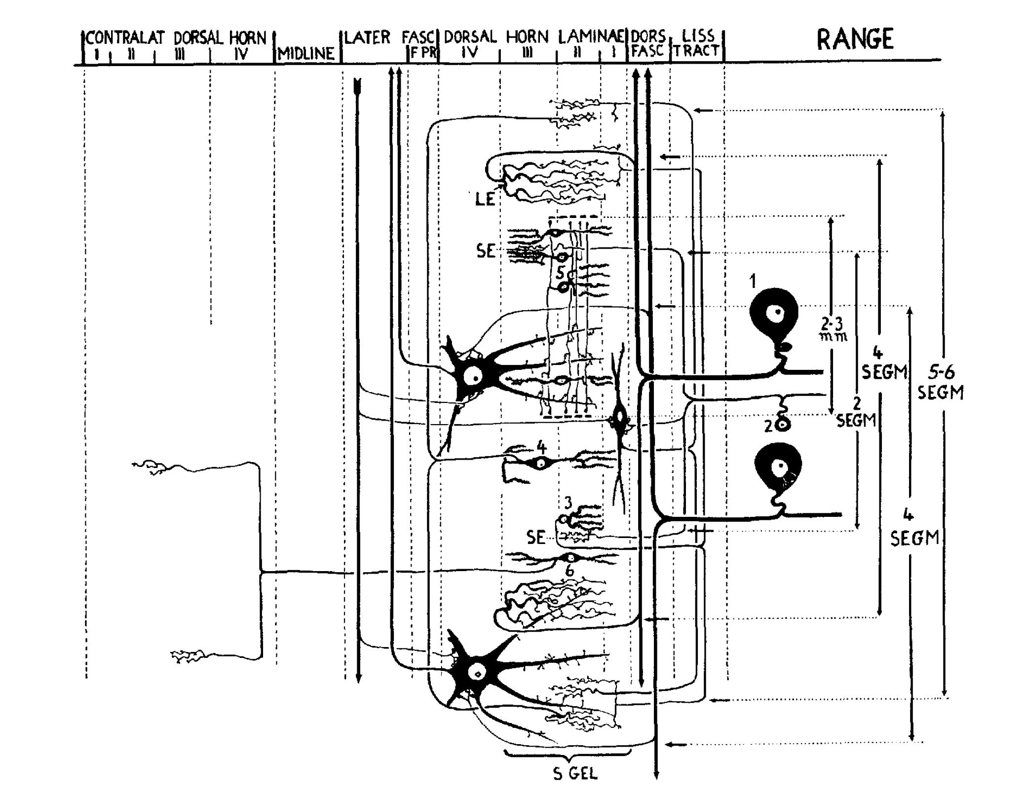
Highly schematized diagram showing neuronal connections of the SG in an imaginary longitudinal section plane that runs through all relevant structures, to illustrate maximal distances bridged. The tracts of the white and the layers of the gray matter are indicated above, the ranges of the several longitudinal connections are indicated at the right side. All other numbers, and labels refer to the same elements as indicated in figure.